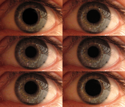
Laser Eye Surgery
 The structure of the eye is composed of a single, outwardly curved (convex) clear lens, the cornea, at the front and a lengthy 'fiber optic' cable, the optic nerve, extending from the back.
It is essentially an empty structure, except for the colored iris, a circular band of muscles that controls the size of the pupil, which allows variable amounts of light to pass to the back inside the surface of the eye. The amount of pigmentation of the iris determines its color. Blue eyes, for example, have very little amount of pigment, and black eyes have the most.
The pupil is the central transparent area, that is controlled by the ciliary muscles in the iris, that make the pupil smaller when the amount of light is excessive, and vice versa.
Light rays pass through the clear cornea, which due to its curved surface, is able to bend (refract) the light rays. These light rays are concentrated together and pass through the pupil. Then, they go through the normally clear lens which has two curved surfaces, the front and the back. Therefore, these light rays are bent (refracted) two more times on their trip to the back of the eye.
The light rays travel to the back surface of the eye through the vitreous, a clear jelly which fills the space between the back of the lens and the retina, the inside lining of the back surface of the eye which contains specialized cells which convert light energy into electrical impulses.
These cells are either called rods, specialized for black and white images, or cones, that mainly process color images. In dim light, we use our rods, which cannot work in bright light. To deal with bright or moderate light, we use our cones, that beside providing color vision, they also process some aspects of black and white vision and the ability to discern fine detail.
What is truly amazing about the eye is how part of these cells in the retina (photosensitive cells) actually are a six inch appendage of the cell, the axon, which joins with other axons to compose the optic nerve which travels to the brain stem, the very top of the spinal cord, located at the very center of the brain. There, each axon connects (synapses) with a cell or cells, and the axon of the receiving cell(s) travels another six inches to the back of the brain, the occipital lobe, where it synapses with a brain cell(s) to produce what we call vision. Therefore, the major functions of these parts of the visual system are composed by:
- Cornea: Refracts light rays
- Pupil: Controls the amount of light entering the eye
- Lens: Refracts light rays
- Vitreous: Light traverses this space
- Retina: Converts light energy to electrical energy
- Optic Nerve: Transmits electrical energy from the retina to the brain stem
- Brain Stem: Intermediate 'relay station' for visual fibers
- Occipital Cortex: Final destination. Converts electrical energy to visual images
A "Perfect Eye" would therefore have:
- a clear and unobstructed path from the front of the eye to the back of the eye.
- the proper balance between the length of the eye and the curvatures of the three refracting surfaces.
- properly functioning cells in the retina and brain which allow the conversion of light energy to electrical energy, the transmission of this energy, and the interpretation of the energy into what we call vision.
Eyes that are too long or have too much refracting power (from the cornea and the lens) are nearsighted eyes, as images are focused in front of the retina. The image received by the retina is not a 'dot for dot' representation of what the image viewed by the eye. Instead, each of these 'dots' of light becomes enlarged to form a 'disc' of light with a consequent spread of the dot image to adjacent parts of the retina. This is what causes blurring of vision.
The opposite results when eyes or too short or have too little refracting poser. These eyes are farsighted, as images are focused (or would be) behind the retina. The same type of dot to disc representation occurs.
When light rays that are vertically oriented are not refracted the same amount as the light rays that are horizontally oriented, this condition is called astigmatism. An example would be when that eye looks at a building that is built as a square, it would appear as a rectangle with different vertical and horizontal dimensions being visualized. This example refers to strictly vertical (90 degrees) and strictly horizontal (0 degrees); astigmatism can occur at any angle between 0-180 degrees.
On a more dynamic level, the eye can possess no refractive error whatsoever but is unable to adjust to near images by increasing its refractive powers. This condition is termed presbyopia and normally affects persons in their 40s and early 50s. The mechanisms within the eye that can increase the curvature of the lens (accommodation) become less efficient and external plus lenses are necessary to bring the near images into focus.
The eye is truly like a camera because it not only requires proper focusing (refraction) but it requires a clear media through which light rays must pass. Any loss of clarity of the structures through which these light rays must pass will interfere with their successful interpretation within the visual cortex of the brain. Examples of disorders which might cause this scattering or absorption of light rays are opacities or swelling of the cornea (scars, edema, abrasions, etc.), opacification of the lens (cataract), and cloudiness of the vitreous (hemorrhage or inflammation.)
Also, the receiving tissue, the retina must be functioning properly, as opposed to aged related macular degeneration, in which there is deterioration of the most important part of the retina, the macula, which is responsible for our finest and our reading vision.
The range of patologies which can be treated with Laser Eye Surgery are:
- Myopia (nearsightedness): -0.75 diopters to -10.0 diopters
- Hyperopia (farsightedness): +0.75 diopters to +5.0 diopters
- Astigmatism (associated with myopia): 0 to 4.0 diopters
- Presbyopia (the eye's diminished ability to focus that occurs with aging).
There are three main types of Laser Eye Surgery:
PRK (Photo Refractive Keratectomy) has been the standard eye laser surgery for many years.
It involves the complete removal of the epithelium or the outer layer of cells on the eye, with the use of a "cold" laser light.
PRK is mostly used on patients with low to moderate amounts of Myopia Astigmatism.
Below you can watch a video showing a PRK surgical intervention:
LASEK (Laser Assisted Sub-Epithelial Keratectomy) or EPIFLAP (Laser Assisted Epithelial Keratomileusis) surgery involves the fractional removal of the epithelium to reveal the cornea, which is then firmed or reshaped by a laser. The outer layer of cells is then pushed back, and the eye heals shortly after.
LASEK is used on patients with low to moderate amounts of Myopia or Astigmatism.
Below you can watch a video showing a LASEK surgical intervention:
LASIK (Laser Assisted in Situ Keratomileusis) is the newest and most popular type of eye laser surgery. Initially developed by Spanish ophthalmologist José Barraquer, with this procedure a thin layer of the eye's cornea is sliced off to create a flap. This enables the laser to target the tissue underneath. The flap is simply replaced and grows back naturally. Vision is restored almost immediately.
LASIK is usually used on patients with severe Myopia (nearsightedness): -0.75 diopters to 10.0 diopters.
Below you can watch a video showing a LASIK surgical intervention:
All of these treatments have been approved for use in many countries around the world and are therefore considered to be safe.
But it must be taken into account that all types of surgery does involve risks and eye surgery is no different.
There have been cases during which patients have suffered major complications and required cornea transplants following laser treatment.
Not everyone can have laser eye surgery. Although eye laser surgery is usually suitable for healthy adults and can be performed on virtually any eye, sometimes it is not advisable.
Several factors could influence negatively the outcome of these operations:
AGE: It is advisable that a person undergoing Laser Eye Surgery should be at least eighteen years old. It is assumed that the refractive error (prescription) of the patient will be fairly stable at this time.
However, there's no upper age limit for Laser Eye Surgery eligibility, since it does not affect the nature or outcome of the surgery.
ENDOCRINE: Hormone fluctuations can affect the refractive error of a person's eye. Surgery during a period of such instability would probably produce changes which would not be correct once the endocrine system returns to normal. Therefore, Laser Eye Surgery is not recommended for women who may be pregnant, nursing, or undergoing changes in oral contraceptive therapy.
HEALTH: Because the cornea is composed of a matrix of collagen fibers, the same material which comprises tendons and other connective tissue, people who have autoimmune disorders, e.g., rheumatoid arthritis, Sjogren's Syndrome, Systemic Lupus Erythematosis, etc., may be at risk for unpredicted changes following surgery. Other medical problems, such as diabetes mellitus, high blood pressure, heart problems, etc. do not affect the outcome of Laser Eye Surgery surgery.
HERPES VIRUS INFECTION: A history of herpes simplex infection of the eye may be a contraindication to Laser Eye Surgery surgery. It is believed that the virus may permanently reside in nerve cells outside the eye and that any kind of surgery might reactivate an infection. However, this area is undergoing change. Studies are being done to see if treatment with antiviral medication prior and after the operation might prevent such a recurrence. Herpes infections in other parts of the body do not pertain to qualifying for Laser Eye Surgery surgery.
PUPIL SIZE: The excimer lasers currently treat an area of the cornea that measures about 7 to 8 mm in diameter. Therefore, it is important that the pupil size be less than the area of treatment. Most of the reported difficulties with glare experienced by patients come from those with pupillary size irregularities.
REFRACTIVE ERROR: Most persons are either nearsighted or farsighted with some degree of astigmatism. After age of 40, people also experienced difficulties with focusing at near, a condition known as presbyopia.
Although none of the procedures described above can be used for this condition, for such patients there exists laser thermal keratoplasty (LAPR), in which mild laser heat is used in a three-second procedure to shrink collagen in the periphery of the cornea to steepen the eye's surface for correction of near vision in farsightedness or presbyopia.
End results are similar to Conductive Keratoplasty (CK), which is also used to treat this condition. This procedure uses radio frequency energy to apply heat to very tiny spots around the cornea.
External links


4 comments:
Fantastic Article! I did not know that there were system for mapping a persons individual eyes. While doing my research I came across "WaveScan WaveFront" which does just that!
LASIK is a surgical procedure that uses a laser to correct nearsightedness, farsightedness, and/or astigmatism. In LASIK, a thin flap in the cornea is created using either a microkeratome blade or a femtosecond laser. The surgeon folds back the flap, then removes some corneal tissue underneath using an excimer laser. The flap is then laid back in place, covering the area where the corneal tissue was removed.
A very good article.. which clearly explains the the pros and cons of the LASIK surgery
.Thank you for the keen information and i am happy that you share this information in very simple language and very efficiently.I came to know many things about our eyes today.
Can we use Contact Lenses after Lasik Eye Surgery. ?
Post a Comment